Breast biopsy
Definition
A breast biopsy is the removal of breast tissue for examination by a pathologist. This can be accomplished surgically or by extracting, or withdrawing, tissue through a needle.
Purpose
A biopsy is recommended when a significant abnormality is found by physical examination or an imaging test. Examples of an abnormality can include a breast
Demographics
The American Cancer Society estimated that in 2003, 211,300 new cases of breast cancer would be diagnosed in the United States and 39,800 women would die as a result of breast cancer. Approximately one in eight women will develop breast cancer at some point in her life. The risk of developing breast cancer increases with age: women ages 30–40 have a one-in-252 chance, women ages 40–50 a one-in-68 chance, women ages 50–60 a one-in-35 chance, and women ages 60–70 a one-in-27 chance.
In the 1990s, the incidence of breast cancer was higher among Caucasian women (113.1 cases per 100,000 women) than African American women (100.3 per 100,000). The death rate associated with breast cancer, however, was higher among African American women (29.6 per 100,000) than Caucasian women (22.2 per 100,000). Death rates were lower among Hispanic women (14.2 per 100,000), Native American women (12.0), and Asian women (11.2 per 100,000).
Description
The type of biopsy recommended will depend on whether the area can be felt, how well it can be seen on mammogram or ultrasound, and how suspicious it feels or appears. Specialized equipment is needed for different types of biopsy and availability may vary.
Surgical biopsy
There are two major types of surgical breast biopsy: excisional and incisional. An excisional biopsy is a surgical procedure where the entire area of concern and some surrounding tissue is removed. It is usually done as an outpatient procedure in a hospital or freestanding surgery center. The patient may be awake and is sometimes given medication to make her drowsy. The area to be operated on is numbed with local anesthetic. Infrequently, general anesthesia is used. An excisional biopsy itself usually takes under one hour to perform. The total amount of time spent at the facility depends on the type of anesthesia used, whether a needle localization was done, and the extent of the surgery.
If a mass is very large, an incisional biopsy may be performed. In this case, only a portion of the area is removed and sent for analysis. The procedure is the same as an excisional biopsy in other respects.
Needle biopsy
A needle biopsy removes part of the suspicious area for examination. There are two types: aspiration biopsy (using a fine needle) and large-core needle biopsy. Either of these may be called a percutaneous needle biopsy. Percutaneous refers to a procedure done through the skin.
A fine-needle aspiration biopsy uses a very thin needle to withdraw fluid and cells that can be studied. It can be done in a doctor's office, clinic, or hospital. Local anesthetic may be used, but is sometimes withheld, as its administration may be more painful than the biopsy needle. The area to place the needle may be located by touch without using specialized equipment. However, ultrasound guidance enables the physician to feel and see the lesion at the same time. The actual withdrawal of fluid and cells can be visualized as it occurs. This helps ensure that the specimen is taken from the right place.
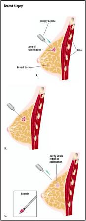
A large-core needle biopsy uses a larger diameter needle to remove small pieces of tissue, usually about the size of a grain of rice. It can be done in a clinic or hospital that has the appropriate facilities. Local anesthetic is routinely used. Ultrasound or x ray is used for guidance of a large-core needle biopsy.
If the suspicious area is seen best with x ray, a stereotactic device is used. This means that x rays are taken from several angles. The information is fed into a computer that analyzes the data and guides the needle to the correct place. The patient may be sitting up, or she may be lying on her stomach, with her breast positioned through an opening in the table. The breast is held firmly but comfortably between a plastic paddle and a metal plate, similar to those used for mammograms. X rays may be taken before, during, and after the tissue is drawn into the needle to confirm that the correct spot is biopsied. This procedure may also be referred to as a stereotactic core biopsy, or a mammotomy.
Ultrasound is used to guide needle placement for some lesions. The patient lies on her back or side. After the area is numbed, sterile gel is applied. The physician places a transducer, an instrument about the size of an electric shaver, over the skin. This produces an image from the reflection of sound waves. A special needle, usually in a spring-loaded device, is used to obtain the tissue. The procedure is observed on a monitor as it is happening.
An abnormal pathology report indicates a cancer is present. If a fine-needle aspiration biopsy was performed, the pathologist has viewed individual cells under a microscope to see if they appear cancerous. Large-core needle biopsy and surgical biopsy will be able to give more information. This includes the type of cancer, whether or not it has invaded surrounding tissue, and how likely it is to spread quickly. There are some conditions that are not malignant but indicate high risk for future development of breast cancer. If these are identified, more frequent monitoring of the area may be recommended.
Diagnosis/Preparation
Sometimes an abnormality can be palpated during a self-examination or an examination by a health care professional. If an abnormality is not felt, there are other signs that indicate the need for medical attention. These include:
- severe breast pain
- changes in the size of a breast or nipple
- changes in the shape of both breast and nipple
- pitting, dumpling, or redness of the breast skin
- nipple redness, irritation, or inversion
- changes in the pattern of veins visible on the surface of the breast
- some types of nipple discharge
If the abnormality cannot be located easily, a wire localization may be done before the actual surgery. After local anesthetic is administered, a fine wire is placed in the area of concern. Either x ray or ultrasound guidance is used. The wire can then be followed to the area of concern. The patient is awake and usually sitting up.
A surgical breast biopsy may require the patient to have nothing to eat or drink for a period of time before the operation. This will typically be from midnight the night before, if general anesthesia is planned. No food restrictions are necessary for needle biopsy, although it is advisable to eat lightly before the procedure. This is especially important if the patient will be lying on her stomach for a stereotactic biopsy.
Aftercare
After a surgical biopsy, the incision will be closed with stitches and covered with a bandage. The bandage can usually be removed in one or two days. Stitches are taken out approximately one week afterward. Depending on the extent of the operation, normal activities can be resumed in approximately one to three days. Vigorous exercise may be limited for one to three weeks.
The skin opening for a needle biopsy is minimal. It may be closed with thin, clear tape (called a steri-strip) or covered with a small bandage. The patient can return to her usual routine immediately after the biopsy. Strenuous activity or heavy lifting is not recommended for 24 hours. Any bandages can be removed one or two days after the biopsy.
Risks
Infection is always a possibility when the skin is broken, although this rarely occurs. Redness, swelling, or severe pain at the biopsy site would indicate a possible infection. Another possible consequence of a breast biopsy is a hematoma. This is a collection of blood at the biopsy site; the body usually absorbs blood naturally. If the hematoma is very large and uncomfortable, it may need to be drained. A surgical breast biopsy may produce a visible scar on the breast, which may make future mammograms harder to interpret accurately.
A false negative pathology report is another risk. This means that no cancer was found when cancer was actually present. The incidence of this varies with the biopsy technique. In general, fine-needle aspiration biopsies have the highest rate of false negative results, but there may be variation in results between facilities.
Normal results
A normal pathology report indicates no malignancy is present. The tissue sample may be further classified as a benign breast condition, including tumor of the breast (fibroadenoma) and connective tissue that resembles fiber (fibrosis). Studies have demonstrated that approximately 80% of all breast biopsies result in a benign pathology report.
Morbidity and mortality rates
The reported rate of complications for image-guided percutaneous biopsy ranges is approximately 2%. Excessive bleeding occurs after approximately 0.5% of fine needle biopsies, 3% of small needle biopsies, and 5% to 10% of large needle biopsies. Infection occurs in approximately 1% of biopsy sites. Organ damage such as a collapsed lung (pneumothorax) occurs in approximately 0.5% of biopsies.
Alternatives
While a biopsy is the only way to determine definitively if a breast abnormality is cancerous, there are a number of procedures that may be used to rule out cancer so that a biopsy is not necessary. These include mammography , ultrasound imaging, and ductography (used for imaging the breast ducts and diagnosing the cause of abnormal nipple discharges).
Resources
periodicals
Centers for Disease Control and Prevention. "Recent Trends in Mortality Rates for Four Major Cancers, by Sex and Race/Ethnicity." Morbidity and Mortality Weekly Report, 51, no. 3 (January 25, 2002): 49–53.
Marks, James S. and Nancy C. Lee. "Implementing Recommendations for the Early Detection of Breast and Cervical Cancer Among Low-income Women." Morbidity and Mortality Weekly Report, 49, no. RR02 (March 31, 2000): 35–55.
organizations
American Cancer Society. 1599 Clifton Rd., NE, Atlanta, GA 30329-4251. (800) 227-2345. http://www.cancer.org .
National Cancer Institute. Building 31, Room 10A31, 31 Center Drive, MSC 2580, Bethesda, MD 20892-2580. (800) 422-6237. http://www.nci.nih.gov .
other
Cardella, John F., et al. "Quality Improvement Guidelines for
Image-Guided Percutaneous Biopsy in Adults." Society of Cardiovascular and Interventional Radiology. November/December 1996 [cited March 11, 2003]. http://www.scvir.org/clinical/T26.htm .
"How is Breast Cancer Diagnosed?" American Cancer Society. 2003 [cited March 11, 2003]. http://www.cancer.org/docroot/CRI/content/CRI_2_4_3X_How_is_breast_cancer_diagnosed_5.asp .
"Lifetime Probability of Breast Cancer in American Women." National Cancer Institute. September 13, 2002 [cited March 11, 2003]. http://cis.nci.nih.gov/fact/5_6.htm .
Ellen S. Weber, MSN Stephanie Dionne Sherk
WHO PERFORMS THE PROCEDURE AND WHERE IS IT PERFORMED?
The breast biopsy is usually performed by a surgeon or a radiologist, a medical doctor who specializes in the use of imaging techniques for diagnosis or treatment. The extracted tissue samples are analyzed by a pathologist, a medical doctor who has completed specialized training in the diagnosis of diseases from microscopic analysis of cells and tissues. Surgical biopsies are generally performed at a hospital or surgery center. Because needle biopsies are less invasive, they may be performed at a doctor's office, clinic, or hospital.
QUESTIONS TO ASK THE DOCTOR
- Why is a biopsy recommended?
- What type of biopsy will I have?
- How long will the procedure take?
- When will I find out the results?
- What will happen if the results are positive for cancer?
She is scheduled to meet with a surgeon ref biopsy.
My wife & I both understand the "mechanics" of various biopsy procedures. We are more interested in the results of excisional vs aspiration vs large bore needle biopsy. Her lesion is small (
Too many false positive results from poor and questionably-performed imaging are not only resulting in altering the normal functioning of the body, but reducing the quality of life to an ongoing cancer-hunt - even if there is no malignancy found.
I am now bewildered has anyone ever had this happen?
I had mammograms and then core suction biopsy-followed by 2 more suction core biopsies
Now am going for open surgical biopsy
All because of tiny calcifications which may never become cancerous
My quality of life has gone- still in pain since 1st Aug
Have 2 metal pins in also
May never find cancer and am disfigured and lost quality of life for nothing
Wish I have never had mammograms